Knee
Knee Anatomy
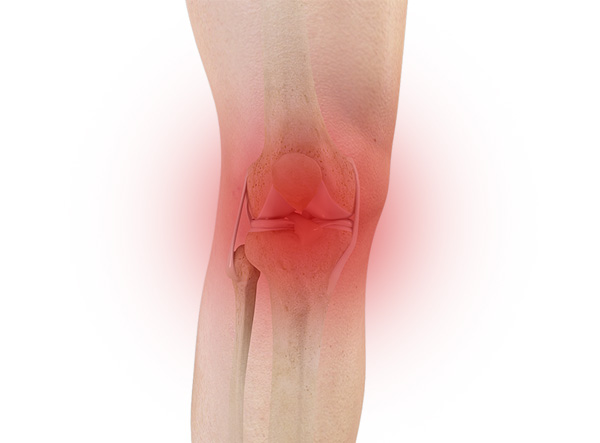
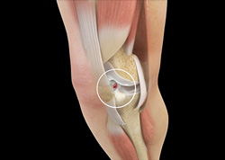
The knee is a complex joint made up of different structures including bones, tendons, ligaments and muscles. They all work together to maintain normal function and provide stability to the knee during movement.
Having a well-functioning healthy knee is essential for your mobility and ability to participate in various activities. Understanding the anatomy of the knee enhances your ability to discuss and choose the right treatment procedure for knee problems with Dr. Fischer.
Bones
The Knee is a large joint made up of two bones, the thigh bone (femur) and the shinbone (tibia). There are two round knobs at the end of the femur called femoral condyles which articulate with the flat surface of the tibia called the tibial plateau. The tibia plateau on the inside of the leg is called the medial tibial plateau, and on the outside of the leg it is called the lateral tibial plateau.
The two femoral condyles form a groove on the front (anterior) side of the knee called the patellofemoral groove, or trochlea. A small bone called the kneecap (patella) articulates with this groove as part of the extensor mechanism. It acts to enhance the knees extension power and efficiency.
A fourth bone called the fibula is the other bone of the lower leg. This form a small joint with the tibia. This joint has very little movement and is not considered a part of the main joint of the knee.
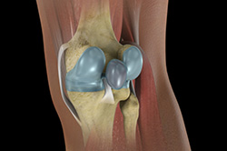
Articular Cartilage & Menisci
Movement of the bones causes friction between the articulating surfaces. To reduce this friction, all articulating surfaces involved in movement are covered with a white, shiny, slippery layer called articular cartilage. The articulating surface of the femoral condyles, tibial plateau and the back of the patella are covered with this cartilage. The cartilage provides a smooth surface that facilitates easy movement.
To further reduce friction between the articulating surfaces of the bones, the knee joint is lined by a synovial membrane which produces a thick clear fluid called synovial fluid. This fluid lubricates and nourishes the cartilage and bones inside the joint capsule.
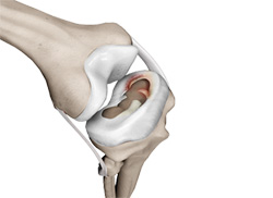
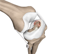
Within the knee joint between the femur and tibia there are two C shaped cartilaginous structures called menisci. Menisci function to provide stability to the knee by spreading the weight of the body evenly across the surface of the tibial plateau. The menisci help in load bearing by preventing the weight from concentrating onto a small area, which could overload the articular cartilage. The menisci also act as a cushion between the femur and tibia by absorbing the shock produced by activities such as walking, running and jumping.
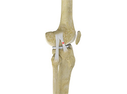
Ligaments
Ligaments are tough bands of tissue that connect one bone to another bone. The ligaments of the knee function to stabilize the knee joint. There are two important groups of ligaments that hold the bones of the knee joint together, collateral ligaments and the cruciate ligament.
Collateral ligaments are present on either side of the knee. They function to prevent the knee from moving too far during side to side motion. The collateral ligament on the inside is called the medial collateral ligament (MCL) and the collateral ligament on the outside is called the lateral collateral ligament (LCL).
Cruciate ligaments - This group of ligaments, present inside the knee joint, control the back and forth motion of the knee. The anterior cruciate ligament (ACL) prevents excess anterior tibial translation and the posterior cruciate ligament (PCL) prevents excess posterior tibial translation.
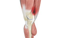
Muscles
Muscles: There are three major muscle groups that power the knee. The quadricep muscles are in the front of the thigh. When the quadriceps muscles contract, the knee straightens. The hamstrings are in the back of the thigh and the gastrocnemius is in the back of the leg. When the hamstring and gastrocnemius muscles contract, the knee bends.
Tendons
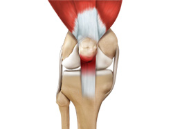
Tendons are structures that attach muscles to the bone. The quadriceps muscles of the knee meet just above the patella and attach to it through a tendon called the quadriceps tendon. The patella further attaches to the tibia through a tendon called the patella tendon. The quadriceps muscle, quadriceps tendon and patellar tendon all work together to straighten the knee. This unit is called the extensor mechanism. Similarly, the hamstrings and gastrocnemius behind the knee power knee flexion.
Understanding Knee Pain
Osteoarthritis :: Rheumatoid Arthritis :: Post-Traumatic Arthritis
Osteoarthritis
Osteoarthritis (OA) is sometimes called degenerative arthritis because it comes from inflammation of the joint due to "wear and tear". Joint cartilage is a smooth covering of the bone ends where they come into contact within the joint. When cartilage wears away, the bones rub against each other, causing pain and stiffness. OA usually occurs in people over age 50 and often in people with a family history of the disease.
Osteoarthritis is the most common form of arthritis in the United States. When OA begins to affect a joint, a series of reactions take place that actually begin to degrade your once-healthy cartilage. Once the cartilage that normally cushions the joint breaks down, the bones of your joint eventually grind directly against each other. Your body reacts to this by creating bone spurs and the joint capsule itself may thicken and become inflamed.
There are two kinds of OA. Primary OA often refers to “everyday wear”. Secondary OA is considered the result of a maligned joint, being overweight, injury or overuse.
Who suffers from OA?
Twenty-seven million people are affected by OA in the United States alone. Although OA can affect anyone at any age, it has been linked to the aging process. More than 50% of everyone over 65 has OA symptoms in one or both knees. By 75, virtually everyone suffers with OA in one or more joints. In fact, OA of the knee and hips continues to be the most common cause of arthritis-related disability for Americans.
What are the Symptoms?
Although some people who have OA say they feel no pain, most people who have OA experience pain, stiffness (especially in the morning), swelling, grinding and tenderness in one or more joints. For some people, OA can become completely debilitating. In order to diagnose you properly, Dr. Fischer will consider your symptoms, medical history, examine your joint(s) and order diagnostic tests. Dr. Fischer may order blood work, X-rays, a CT scan or an MRI to get a better understanding of your painful joint and its condition.
Rheumatoid Arthritis
Rheumatoid arthritis (RA) is an autoimmune disease in which the body’s natural immune response attacks the lining of the joints (called the synovium), causing chronic inflammation and pain. The inflammation may eventually damage the joint’s cartilage and bone, weaken the soft tissue around the joint (cartilage, ligaments and tendons) and prevent the joint from working properly.
Left untreated, RA can greatly reduce your quality of life. You may have already decreased your activity level to avoid the pain caused by a joint affected by RA. It’s not uncommon for the joint damage caused by RA to lead to a loss of movement, an inability to work, and even the need for surgery to repair the damage.
What Are The Symptoms of RA?
RA is a chronic, persistent disease that can take a variable course over an affected person’s lifetime. It may progress slowly, sometimes produce “flare ups”, and then at times go into “remission” during which the symptoms may greatly diminish or disappear. There are 3 stages of RA identified by specific symptoms. In the first stage, RA causes pain, warmth, redness and swelling in affected joints. In the second stage, it causes thickening of the synovium. In the third stage, permanent joint damage begins to occur as bone and cartilage are attacked by the enzymes released by the inflamed cells in the once-healthy synovial fluid.
In addition to joint pain, swelling and stiffness, the symptoms of RA commonly include fatigue, weakness, flu-like symptoms accompanied by a low-grade fever, loss of appetite, depression, chronic dry eye or dry mouth and, in people with more advanced RA, bumps (called rheumatoid nodules) under the skin.
How is RA Treated?
Your primary doctor will refer you to a rheumatologist, a doctor who specializes in inflammatory diseases like RA. Your rheumatologist may recommend different treatment options depending on the severity of your RA and its impact on your joint(s) and your body as a whole. And while there is no cure, RA can be controlled through the use of new drugs, exercise, and self-management techniques.
If your RA is so severe that conservative treatment options are no longer managing your symptoms to meet your activity goals, joint replacement may be an option. Some treatment options, such as partial knee replacement, are not indicated for patients with RA because the underlying disorder will inevitably affect the other native parts of the knee. Dr. Fischer will work with you and your other physicians to develop an individual treatment plan
Post-Traumatic Arthritis
What Are The Symptoms of Post-Traumatic Arthritis?
The symptoms of post-traumatic arthritis include joint pain, swelling, fluid accumulation in the joint, and decreased tolerance for walking, sports, stairs and other activities which stress the joint.
How is Post-Traumatic Arthritis Treated?
Post-traumatic arthritis is treated similarly to osteoarthritis. Treatment starts with weight loss, low impact exercise and strengthening of the muscles surrounding the joint, non-steroidal anti-inflammatory medicines (NSAIDs such as Advil, Aleve, Celebrex or one of many others) are often recommended if you can take them. Arthritic joints can also be injected with cortisone or substances called Hylamers which act like artificial joint fluid. All of these measures are aimed at making the joint more comfortable and functional. They do not cure the arthritis.When the arthritis progresses to the point that these measures are not effective in treating pain and maintaining function, then surgical treatment will be discussed.
Surgical treatment may include debriding ("cleaning out"), reconstructing or replacing the worn out joint surfaces. Post-traumatic arthritis progresses as time goes on, the joint surface wearing out further with more use over the years. Fortunately, when the nonsurgical treatments are no longer effective, surgical treatment can offer lasting relief. Deformity through or around the joint may necessitate the use of computer assisted navigation to restore the joints normal alignment.
Treating Knee Pain
Dr. Fischer may recommend different treatment options depending on the severity of your arthritis and your specific activity goals.
Treatment starts with weight loss, low impact exercise and strengthening of the muscles surrounding the joint, non-steroidal anti-inflammatory medicines (NSAIDs such as Advil, Aleve, Celebrex or one of many others) are often recommended if you can take them. Arthritic joints can also be injected with cortisone or substances called Hylamers which act like artificial joint fluid. All of these measures are aimed at making the joint more comfortable and functional. They do not cure the arthritis. When the arthritis progresses to the point that these measures are not effective in treating pain and maintaining function, then surgical treatment will be discussed.
Dr. Fischer will determine the proper surgical treatment based on the severity of your arthritis and its location. Today, a full range of surgical solutions exist that enable Dr. Fischer to tailor the treatment to your particular needs and anatomy.
Surgical Treatment Options
Conditions
Knee Pain
The knee is one of the largest joints in the body, formed by the lower end of the femur, upper end of the tibia and the patella or knee cap. Several ligaments and muscles attach to the bones of the knee joint to maintain normal motion of the joint. Special cartilaginous tissues known as menisci are placed between the two articular ends of the joint. These act as a cushion between the articular surfaces and absorb the shock during movement.

Anterior Knee Pain
Anterior knee pain is a characterized by a chronic pain over the front and center of the knee joint. It is common in athletes, active adolescents (especially girls) and overweight individuals. Anterior knee pain refers to a variety of conditions which include runner's knee or patellar tendinitis and chondromalacia of the patella.
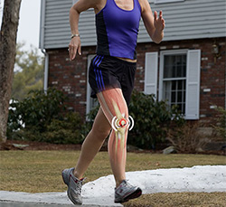
Runner’s Knee
Runner's knee, also called patellofemoral pain syndrome refers to pain under and around your kneecap. Runner’s knee includes several medical conditions such as anterior knee pain syndrome, patellofemoral malalignment, and chondromalacia patella that cause pain around the front of the knee.
Chondromalacia Patella
Chondromalacia patella is a common condition characterized by softening, weakening, and damage of the cartilage. The condition is most often seen among young athletes and older adults who have arthritis of the knee. It is especially seen in women.
Jumper’s Knee
Jumper’s knee, also known as “patellar tendinitis" is an inflammation of the patellar tendon that connects your kneecap (patella) to your shinbone. This tendon helps in extension of the lower leg.
Bursitis
A bursa is a small fluid-filled sac found between soft tissues and bones. It lubricates and acts as a cushion to decrease friction between bones when they move. Bursitis refers to the inflammation and swelling of the bursa. Inflammation of the bursa in front of the kneecap (patella) is known as kneecap bursitis or prepatellar bursitis.
Baker’s Cyst
The knee consists of a fluid called synovial fluid, which reduces friction between the bones of the knee joint while you move your leg. Sometimes this fluid is produced in excess, resulting in its accumulation in the back of your knee. A Baker’s cyst or popliteal cyst is a fluid-filled swelling that develops into a lump behind the knee. This causes stiffness, tightness and pain behind your knee. It is commonly seen in women and people aged over 40 (although it can develop at any age).
Iliotibial Band Syndrome
Iliotibial band syndrome is an overuse injury resulting from the inflammation of iliotibial band. Iliotibial band is a tough group of fibers that begins at the iliac crest of hip and runs along the outside of the thigh, to get attached to the outer side of the shin bone just below the knee joint. Its function is to coordinate with the thigh muscles and provide stability the knee joint.
Osteochondritis Dissecans
Osteochondritis dissecans is a joint condition in which a piece of cartilage, along with a thin layer of the bone separates from the end of the bone because of inadequate blood supply. The separated fragments are sometimes called “joint mice”. These fragments may be localized, or may detach and fall into the joint space causing pain and joint instability.
Knee Injury
Pain, swelling and stiffness are the common symptoms of any damage or injury to the knee. If care is not taken during the initial phases of injury, it may lead to joint damage that may end up destroying your knee.
Unstable Knee
The knee joint is one of the largest joints in the body. This highly complex joint has several tissues supporting and stabilizing its movement:
Knee Sprain
Knee sprain is a common injury that occurs from overstretching of the ligaments that support the knee joint. A knee sprain occurs when the knee ligaments are twisted or turned beyond its normal range causing the ligaments to tear.
ACL Tears
The anterior cruciate ligament, or ACL, is one of the major ligaments of the knee that is in the middle of the knee and runs from the femur (thigh bone) to the tibia (shin bone). It prevents the tibia from sliding out in front of the femur. Together with posterior cruciate ligament (PCL) it provides rotational stability to the knee.
MCL Tears
The medial collateral ligament (MCL) is the ligament that is located on the inner part of the knee joint. It runs from the femur (thighbone) to the top of the tibia (shinbone) and helps in stabilizing the knee. Medial collateral ligament (MCL) injury can result in a stretch, partial tear, or complete tear of the ligament.
Meniscal Injuries
The knee is one of the most complex and largest joint in the body, and is more susceptible to injury. Meniscal tears are one among the common injuries to the knee joint. It can occur at any age, but are more common in athletes playing contact sports.
Meniscal Tears
Knee arthroscopy is commonly recommended surgical procedure for meniscal tears. The surgical treatment options include meniscus removal (meniscectomy), meniscus repair, and meniscus replacement. During meniscectomy, instruments about the size of a pencil are used to remove the torn meniscus. In arthroscopic meniscus repair the torn meniscus will be sutured “all inside” or partially open depending on the extent and pattern of the tear.
Knee Arthritis
Arthritis is a general term covering numerous conditions where the joint surface or cartilage wears out. The joint surface is covered by a smooth articular surface that allows pain free movement in the joint. This surface can wear out for several reasons; often the definite cause is not known.
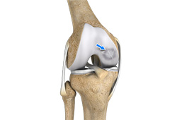
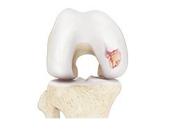
Chondral (Articular Cartilage Defects)
Articular or hyaline cartilage is the tissue lining the surface of the two bones in the knee joint. Cartilage helps the bones move smoothly against each other and can withstand the weight of the body during activities such as running and jumping. Articular cartilage does not have a direct blood supply to it so has less capacity to repair itself. Once the cartilage is torn it will not heal easily and can lead to degeneration of the articular surface, leading to development of osteoarthritis.
Patellar Instability
The patella (kneecap) is a bone in the font of the knee that is part of the extensor mechanism, connecting the quadriceps and patellar tendons. The patella articulates with the femur to form a patellofemoral joint. The patella is protected by ligaments which secure the kneecap from sliding out of place. One of the most important ligaments prevents lateral translation is the medial patellofemoral ligament (MPFL).
Patellofemoral Instability
The knee can be divided into three compartments: patellofemoral, medial and lateral compartment. The patellofemoral compartment is the compartment in the front of the knee between the knee cap and thigh bone. The medial compartment is the area on the inside portion of the knee, and the lateral compartment is the area on the outside portion of the knee joint.
Patella Fracture
The knee cap or patella is the largest sesamoid bone in the body and one of the components of the knee joint, present at the front of the knee. The undersurface of the kneecap and the lower end of the femur are coated with articular cartilage, which helps in smooth movement of the knee joint. The knee cap protects the knee and provides attachment to various muscle groups of the thigh and leg. Fracture of knee cap is rare and is more common in adult males.
Recurrent Patella Dislocation
The patella (knee cap) is a small bone that shields your knee joint. It is found in front of your knee, in a groove called the trochlear groove that sits at the junction of the femur (thighbone) and tibia (shinbone). Articular cartilage present below the patella and end of the femur cushion and help the bones glide smoothly over each other when the legs move.
Quadriceps Tendon Rupture
Quadriceps tendon is a thick tissue located at the top of the kneecap. The quadriceps tendon works together with the quadriceps muscles to allow us to straighten our leg. The quadriceps muscles are the muscles located in front of the thigh.
Patella Tendon Rupture
Patella tendon rupture is the rupture of the tendon that connects the patella (knee cap) to the top portion of the tibia (shin bone). The patellar tendon works together with the quadriceps muscle and the quadriceps tendon to allow your knee to straighten out.
Osteonecrosis of the Knee
Osteonecrosis is a condition in which death of a section of bone occurs because of lack of blood supply to it. It is one of the most common causes of knee pain in older women. Women over the age of 60 years of age are commonly affected, three times more often than men.
Procedures
Non-surgical Treatments
Pharmacological
The knee is a complex joint which consists of bone, cartilage, ligaments and tendons that make joint movements easy and at the same time more susceptible to various kinds of injuries. Knee problems may arise if any of these structures get injured by overuse or suddenly during sports activities.
PRP
Our blood consists of a liquid component known as plasma. It also consists of three main solid components which include the red blood cells (RBCs), white blood cells (WBCs), and platelets. Platelets play an important role in forming blood clots.
Viscosupplementation
Viscosupplementation refers to the injection of a hyaluronan preparation into the joint. Hyaluronan is a natural substance present in the joint fluid that assists in lubrication. It allows smooth movement of the cartilage covered articulating surfaces of the joint.
Steroid Injection
Cortisone is a corticosteroid released by the adrenal gland in response to stress and is a potent anti-inflammatory agent. Artificial preparations containing cortisone are injected directly into the affected joint to relieve pain and reduce inflammation.
Physical Therapy
Physical therapy is an exercise program that helps you to improve movement, relieve pain, encourage blood flow, and restore your physical function and fitness level. The main aim of physical therapy is to make your daily activities such as walking, getting in and out of bed, or climbing stairs easier. It can be prescribed as an individual treatment program or combined with other treatments.
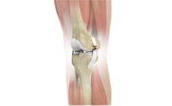
Knee Replacement & Reconstruction
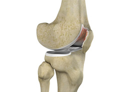
- Unicompartmental Knee Replacement
- Patellofemoral Knee Replacement
- What is new in Knee Replacement
- Total Knee Replacement
- Revision Knee Replacement
- Robotic Assisted Partial Knee Surgery
- Robotic Assisted Knee Replacement
- Outpatient Total Knee Replacement
- Fracture Repair Around a Knee Replacement
Resurfacing
Knee FAQs
How long will I stay in the hospital?
You will likely stay in the hospital for 1 night depending on your rehabilitation protocol and how fast you progress with physical therapy. This is highly dependent upon your condition before surgery, your age, and medical problems which can slow your rehabilitation. Patients who are otherwise healthy are eligible for a rapid recovery and discharge to home the day of surgery.
When can I walk after surgery?
The goal is to get out of bed with physical therapy the day of surgery. Most people are walking with the assistance of a walker on the day after surgery, and using a cane or nothing at all by 2-3 weeks.
How often will I see my surgeon after surgery?
Dr. Fischer is in charge of your care throughout your hospital stay. You can expect to see Dr. Fischer every day while you’re in the hospital recovering. Dr. Fischer will also want to see you for follow-up appointments in his clinic after you are discharged. Follow-up appointments are scheduled 2 weeks after surgery. Further appointments will be made and the timing is based on each individual's needs.
Will I need physical therapy after my knee replacement?
Yes. Physical therapy is an essential part of your recovery after knee replacement. The amount of work you put into your recovery is directly related to the performance of your joint and ultimately your satisfaction with the surgery. Physical therapy is best performed in the outpatient setting. Rarely, therapy may be performed at home with home health.
Is knee replacement very painful?
Pain management following total knee replacement has come a long way with increased use of regional nerve blocks, spinal blocks, and multimodal pain control. Total hip replacement is generally considered to be less painful than total knee replacement. Early range of motion and rapid rehabilitation protocols are also designed to reduce early stiffness and pain, making the procedure in general much less painful than in years past. You may have relatively mild pain following the procedure, or you may have a more difficult time than others. Everyone is unique and handles and perceives pain differently.
Will I need general anesthesia?
While general anesthesia is a safe option, both hip and knee replacements can be performed under regional anesthesia. Choices for regional anesthesia include spinal anesthesia, epidural anesthesia, or one of a variety of peripheral nerve blocks. Many surgeons and anesthesiologists prefer regional anesthesia because data shows it can reduce complications and improve your recovery experience with less pain, less nausea, less narcotic medicine required, etc.
When can I take a shower or bath at home?
Showering (not submerging the wound) is typically allowed immediately after surgery with the water resistant dressing in place. When you return home, you may need special equipment, like a bath mat, hand-held showerhead or shower seat to help you shower comfortably and safely. Taking a bath (submerging the wound) is only allowed after the wound has completely healed over.
When will I be able to drive again?
You should not drive a car or other motor vehicle until Dr. Fischer has approved. You must be off pain medications before you will be cleared to drive again. In most cases, patients are able to resume driving about 4 weeks after surgery.
How should I sleep at night to keep my knee comfortable and safe?
Placing a pillow between your legs should help keep your knee comfortable and stable. You may sleep on your back or on either side, depending on what makes you most comfortable. Placing a pillow underneath your knee is not OK and can result in loss of full knee extension.
How long does it take to recover?
It can take up to 3 months for you to return to most activities, and likely 6 months to one year to fully recover to maximal strength and endurance following a knee replacement. This depends on your condition before surgery, additional medical problems, and your expectations.
How do I know if my incision is infected?
After surgery you will notice some redness, swelling and warmth around your incision. This is normal. If you experience spreading redness, increasing swelling, or persistent drainage from you incision, you may have an infection. A persistent fever greater than 101°F may also indicate infection. A Surgeon is on call 24/7 to address any questions or concerns you may have.
Why must I take antibiotics for dental work or other surgical procedures?
Taking antibiotics is a precaution to help ensure that your new artificial joint does not become infected. Additional surgeries or dental work increases the chance of infection. No matter where the infection starts, if it spreads to your new knee, the results can be serious. When artificial joints become infected, they must be removed surgically and then replaced weeks to months later. Please let your other medical providers know that you have had joint replacement.
What is the lifespan of a knee replacement?
All implants have a life expectancy that depends on several factors including the patient’s weight, activity level, quality of bone stock and compliance with the surgeon’s orders. Proper implant alignment and state of the art materials also contribute to a replaced joint's longevity. With moder bearing materials, a 20-30 year implant life is probable.
Do you use metal on metal implants?
Dr. Fischer does not use Metal on Metal implants for either knee or hip replacements. The knee implants Dr. Fischer uses are state of the art highly cross linked polyethylene (high density plastic) on cobalt chromium (metal) at each point of contact.
What if I have a nickel allergy?
Dr. Fischer has the option to use a nickel free knee replacement that avoids the potential complications to patients who are allergic or sensitive to nickel.